Labs
Research is part of the College's trifold mission of education, research, and patient care. Our research in the Department of Drug Discovery and Biomedical Sciences is concentrated in four areas:
- Bioorganic/Medicinal Chemistry (identification of new drug targets; rational and computer-aided drug design, synthesis, and analysis; drug metabolism; and bioorganic and molecular immunology)
- Pharmaceutics and Pharmacology (microscale and nanoscale drug delivery systems, and ion channel physiology and pharmacology)
- Cell Death, Injury, and Regeneration (mechanisms of drug- and disease-induced cell injury, death and regeneration; molecular and cellular toxicology; drug transport; and acute heart, kidney, liver failure and stroke)
- Cancer Etiology and Therapeutics (mechanisms of cancer chemoprevention; inflammation; genome instability and DNA repair; and cancer therapeutic mechanism of action)
For information about graduate research training, visit Doctor of Philosophy Program Objectives and Requirements
Faculty Labs
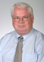
Mark Hamann, Ph.D.
Professor
Charles & Carol Cooper Endowed Chair

Mark Hamann is the Charles and Carol Cooper Chair in Pharmacy Endowed SmartState Chair in the Cancer Drug Discovery Center of Economic Excellence. His team looks at the role of natural products in the discovery and development of therapeutics. It focuses on the discovery and development of new treatments for drug resistant cancer and infectious diseases from natural product prototypes. His group has identified potential new and innovative solutions to pancreatic cancer, leukemia, breast, and lung cancer as well as MRSA and depression.
John J. Lemasters, M.D., Ph.D.
Professor & GlaxoSmithKline
Distinguished Endowed Chair in Cancer Drug Discovery

John LemastersDr. Lemasters’ research interests concern the cellular and molecular mechanisms underlying toxic, hypoxic and ischemia-reperfusion injury to liver and heart. His laboratory applies new techniques of laser scanning confocal and multiphoton microscopy to characterize the physiology of single living cells, including the assessment of ion homeostasis, chelatable iron, mitochondrial function, electrical potentials, oxygen and nitrogen free radical formation, membrane permeability, and other biochemical parameters during the pathogenesis of lethal cell injury. These approaches are being extended to true in vivo (intravital) imaging of cells and tissues in living animals.
Increased Ca2+, formation of reactive oxygen and nitrogen species, and oxidation of pyridine nucleotides and glutathione promote a phenomenon called the mitochondrial permeability transition that in turn leads to mitochondrial depolarization and uncoupling of oxidative phosphorylation. Studies in living cells show MPT initially induces the sequestration and lysosomal degradation of mitochondria by the process of autophagy (mitophagy). However, excess MPT induction induces both necrotic cell death and apoptosis during reperfusion injury, oxidative stress, excitotoxicity, calcium ionophore-induced toxicity, drug toxicity, exposure to tumor necrosis factor-alpha, Fas ligation, and organ preservation for transplantation surgery. Inhibitors of the MPT decrease or abolish cell killing in these models. Progression to apoptosis or necrosis after the MPT depends on the presence or absence, respectively, of ATP. Often, features of both apoptotic and necrotic cell death develop after death signals and toxic stresses. These findings offer new strategies to rescue cells and tissues from irreversible toxic and ischemic injury.
Despite a detailed understanding of their metabolism, mitochondria often behave anomalously. In particular, global suppression of mitochondrial metabolism and metabolite exchange occurs in apoptosis, ischemia and anoxia, cytopathic hypoxia of sepsis and multiple organ failure, alcoholic liver disease, aerobic glycolysis in cancer cells (Warburg effect) and unstimulated pancreatic beta cells. The lab is exploring the hypothesis that closure of voltage-dependent anion channels (VDAC) in the mitochondrial outer membrane accounts for global mitochondrial suppression consistent with a role for VDAC as a dynamic regulator, or governator, of global mitochondrial function both in health and disease.
Anna Liisa Nieminen, Ph.D.
Professor
 The Anna Liisa Nieminen laboratory is investigating mechanisms of cell injury and cell death in various pathological settings, mostly related to cancer. Currently we are interested in treating head and neck cancers with photodynamic therapy.
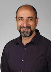
Eduardo N. Maldonado, D.V.M., Ph.D.
Associate Professor

Dr. Maldonado’s laboratory studies mitochondrial metabolism in cancer, cancer bioenergetics and cellular bioenergetics in different cell types. His research program focuses on the interplay between mitochondrial metabolism and the Warburg phenotype in proliferating cells, and the regulation of mitochondrial-dependent bioenergetics in embryonic stem cells, cancer stem cells and skeletal muscle.
A main research line in his lab is the study of the regulation of voltage dependent anion channels (VDACs) located in the mitochondrial outer membrane, as global modulators of mitochondrial metabolism. He recently proposed VDAC opening as a pro-oxidant anti-Warburg switch. The discovery of free tubulin as an endogenous regulator of VDAC opening in cancer cells, led Dr. Maldonado to propose the VDAC-tubulin interaction as a pharmacological target to revert mitochondrial suppression in the Warburg phenotype. The central concept of Dr. Maldonado’s research is that VDAC opening promotes cell killing by a combination of increased oxidative metabolism, oxidative stress and reversal of the Warburg effect. Currently, Dr. Maldonado’s lab is developing small molecules to antagonize the inhibitory effect of tubulin on VDAC and also to stabilize VDAC in an open configuration. The expectation is to develop new drugs for treating liver and other types of cancer that may act as “metabolic modifiers” to be used alone, or as coadjuvants of preexisting therapies.
Overall, Dr. Maldonado’s lab combines basic science approaches including molecular biology, biochemistry and cell biology with drug development. Students, postdocs and scientists at different levels are always welcomed for collaborations, and temporary or long-term visits. Dr. Maldonado also created Mitobonds, a network fostering the worldwide exchange of scientists among labs interested in mitochondrial research.
If you want any additional information or are interested in contacting Dr. Maldonado’s lab, please send an email to maldona@musc.edu.
PubMed Entries
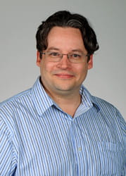
Yuri Karl Peterson, Ph.D.
Associate Professor
Assistant Director of Drug Discovery and Interim Director of Bioenergetics Profiling

Dr. Peterson’s research focus is in applied pharmacologic sciences using in vitro, cell based, and in silico approaches to quantitate protein and small molecule functionality to bridge between chemical biology and pathobiology. He has experience in the experimental biology and modeling of protein-protein interactions, protein-ligand interactions, and hormone signaling. Specifically, his research efforts have included the study of arrestins, the cytoskeleton, GPCRs, heterotrimeric and monomeric G-proteins, scaffolding proteins (like RGS and AGS G-protein regulators), prenyltransferases, methyltransferases, deacetylases, kinases, lipid binding proteins, mitochondria, and endosomes. Highlight innovations from the Peterson group include the discovery and therapeutic utility (Tat-GPR) of guanine nucleotide dissociation inhibitors, software to analyze endosome kinetics (DotQuanta), the discovery of Gi-alpha suppression in the majority of ovarian cancer patients, and methodologies to optimize virtual screening for drug discovery. Dr. Peterson’s current research focus is on the application of high-content microscopy to study cellular protein kinetics and predictive bioinformatics using QSAR, pharmacophores, molecular docking, and machine learning for personalized medicine.
Ed Soltis, Ph.D.
Professor & Vice-Chair for Professional Education
Ed Soltis, Vice-Chair for Professional Education

Prior to his arrival at MUSC in 2002, Dr. Soltis research was focused primarily on the study of vascular smooth muscle physiology and its alterations in hypertension, aging and diabetes using various rodent models. He received national and local support of his research primarily through the NIH, AHA and DREF. He was involved for several years in the oversight and direction of a component of the MUSC College of Graduate Studies Summer Undergraduate Research Program. He currently serves on the MUSC Research Integrity Committee as well as the University Tenure Committee. Dr. Soltis’ current efforts are directed primarily towards teaching and educational initiatives in the College of Pharmacy. He teaches and coordinates courses across all three didactic years of the curriculum and actively participates in various academic and educational committees.
Danyelle Townsend, Ph.D.
Professor

The Townsend lab combines proteomics and drug metabolism with analytical biochemistry to identify molecular targets of oxidative and nitrosative stress and determine how redox signaling impacts cellular response. This research led to the discovery that S-glutathionylation is not a spontaneous redox event, rather it is enzyme-mediated through GSTP activity, which differs in human populations through functional polymorphisms.
We are currently investigating redox signaling pathways that modulate protein disulfide isomerase (PDI) and either trigger or protect from endoplasmic reticulum (ER) stress-induced apoptosis. The ER stress response is a cascade of transcriptional and translational events that sense the folding capacity and attempt to counterbalance ER stress (prosurvival). However, when the ER stress response is unable to handle the protein folding capacity, apoptotic pathways are activated. Unsurprisingly, an accumulation of misfolded proteins is found in a number of human pathologies, including neurodegenerative disorders and cancer. Our laboratory has shown that GSTP-mediated redox regulation of PDI alters its chaperone activity with estrogen receptor alpha (ER). This nodal connection is important to understanding the dual function of PDI in estrogen-responsive pathologies, including Parkinson’s disease and breast cancer.
Thus, the focus of our laboratory is to understand the precursor and/or proximal events leading to stress response, notably through investigating the redox-induced impact on proteins directly involved in the protein folding process.
Zhi Zhong, M.D., Ph.D.
Professor

My research interests are on the mechanisms of cell injury, death and regeneration, with foci on the roles of free radicals and mitochondrial function in liver and kidney diseases, and development of novel therapeutics to prevent injury and accelerate the regeneration/repair processes. In particular, using state-of-the-art intravital confocal/multiphoton microscopy, which allows visualization of cells and organelles in live animals, as well as RNAi and molecular biological techniques in combination with a variety of animal models, isolated organ perfusion and cell culture, we are investigating the mechanisms of mitochondrial dysfunction/biogenesis, the cellular, organelle, and enzymatic resources of free radical production, and the roles of free radicals in alteration of signal transduction that subsequently affect pathogenesis and regenerative processes. We are studying hepatic and renal injury, fibrosis and regenerative processes in alcohol and drug exposure, liver and kidney transplantation, ischemia-reperfusion, obesity, and cholestasis. The overall goal of our research is to gain new insights into the mechanisms and to develop effective therapeutics to prevent and treat these hepatic and renal diseases.
PubMed Entries

